This post is sponsored by a new video analysis app: Athletic Sports Videos
Your muscles help you move and help your body work. Although there is a great amount medical information about the bones and articulations, the knowledge on the muscles role in canine performance is limited. Muscle strength and endurance is studied to a great deal in the human side, related to exercise physiology, conditioning and strength training. I feel there is much to learn in the area of dog muscle physiology and its role in movement and locomotion. After over three decades of studying canine performance, I can say that this area of science is very wide and deep. It cannot be addressed in a simple post. There are a few topics that can be covered in a relatively short space that can be of benefit to those individuals who own, manage, handle or train dogs.
Muscle Activity
Muscles create movement
Muscle Definitions
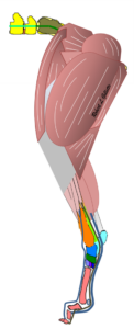
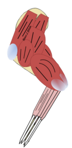
Muscles are the body’s tissues that create motion. They not only create motion, but they also work to stabilize against improper motion. Skeletal muscles can be divided into two groups: Axial Muscles and Appendicular Muscles. Axial muscles are the muscles of the core and include the regions of the head, neck, thorax, abdomen and tail. Appendicular muscles are associated with the limbs. This includes the shoulders, hips and the four legs. When running the energy for locomotion is created by the muscles of the rear legs and lower back. The vertebral muscles work to stabilize the core body. The muscles of the front legs help absorb the energy of impact and work navigate the body.
Appendicular muscles of the front and rear limbs
|
|
 |
Axial Muscles of the vertebral column
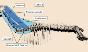 Vertebral Muscles of the Cranial Column |
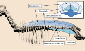 Vertebral Muscles of the Torso |
Some skeletal muscles are considered postural muscles while others are action muscles. In general, the postural muscles act to stabilize the structure and work against gravity. They are typically made up of slow-twitch muscles, which are slow to fatigue. The skeletal (action) muscles work to create movement and locomotion.
Most movement or locomotory actions are learned or trained over time. A puppy is not very agile or does not have concise movement actions. As they mature the nerves and muscles learn to work together to advance the movement capabilities. Muscles grow and adapt to these new actions and body growth. After the neuromusculoskeletal system matures, the body is capable of many movements and actions.
Muscle Components of Action
Locomotion or other body movements occur as a result of balanced and synchronized muscle contractions. Muscles are made up of myofibrils (muscle cells). Muscle cells are composed of millions of fibers and their protein chains. These protein chains (Actin and Myosin) interact with each other to produce muscle contraction.
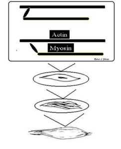
Muscles are composed of muscle cells, which are made of fibers. These fibers are composed of many actin and myosin bands
There is energy needed for these actions. It is provided by ATP. To create energy needed for muscle contraction, a chemical reaction is utilized where ATP is reduced to ADP. This reaction provides the energy that allows the action and myosin chains interact and slide over each other. The actin and myosin bands move along each other to create muscle contractions
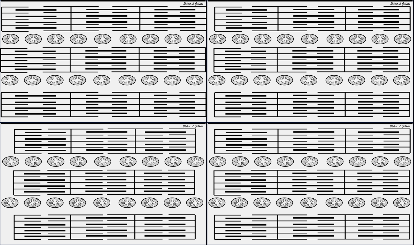
Contraction Energy comes from ATP
ATP Sources
The ATP that is used for muscle contraction needs to be available where the Actin and Myosin (A/M) bands interact. It can come from the immediate energy stores. Some ATP is located within the muscle fibers. Creatine phosphate can bind with ATP and be stored in that location for use when needed. Anaerobic energy, ATP, can be provided from muscle glycogen stores. This can provide quick energy but is not very efficient. Aerobic energy can be created in the mitochondria via the Krebs Cycle (also called the Citric Acid Cycle) and the Electron Transport Chain. These are more complex energy sources but are very efficient.
- Immediate energy stores
- Primary is ATP
- CP can replenish ATP
- Glycolytic Energy
- Muscle Glycogen Stores
- Aerobic Energy
- Mitochondria produce the majority of the ATP used for muscle contraction
- Site of the Kreb’s Cycle and the Electron Transport chain
Myofascial Trigger Points or Taut Muscle Bands
Muscle disorders can be classified as Traumatic Injuries, Inflammation, (is sprains, cramps, myositis, etc), Muscle spasms, Trigger Points, or Medical Disorders (ie muscular dystrophy, cancers, etc).
A myofascial trigger point or trigger point (TP) is where a muscle fiber within the muscle belly is abnormally contracted. It is theorized that certain myofascial situations results in an abnormal accumulation of intracellular Ca2+, persistent A/M cross bridging and then activation of the nociceptive system results in the formation of a trigger point. It is important to note that this is an active contraction of the fibers. It is using ATP to maintain this contraction
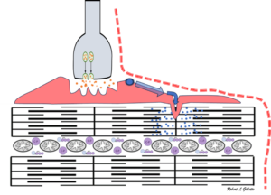
Myofascial Trigger Points & Taut Muscle Bands
A trigger point is where a muscle fiber within the muscle belly is abnormally contracted. It is defined as “a focus of hyperirritability in a tissue, when compressed, is locally tender and if sufficiently hypersensitive, gives rise to referred pain and tenderness, and sometimes to referred autonomic phenomena and distortion of proprioception”. Over time, this area of contraction can expand to where it is affecting almost an entire muscle belly. These are referred to as Taut Muscle Bands (TMB). A TP can occur for many reasons, but they can act in a guarding manner. If there is an incident or event that causes a musculoskeletal injury or fault, they will arise to help minimize the body’s actions that stimulate pain or further injure the tissue. Many times, they will still be present after the primary issue resolves. The trigger point or taut muscle band will act as a guardian mechanism to restrict musculoskeletal actions that are painful or disruptive to the health of the muscles, joints, or bones.
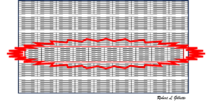 Abnormal fiber contraction within a muscle |
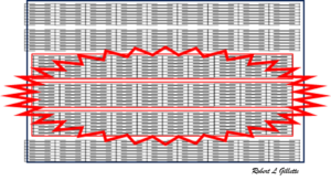 The abnormal contraction expands to surrounding fibers |
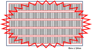 The abnormal fiber contraction can expand across many fibers and may include total muscle tissue |
TPs and TMBs will affect Normal Movement of the Musculoskeletal System
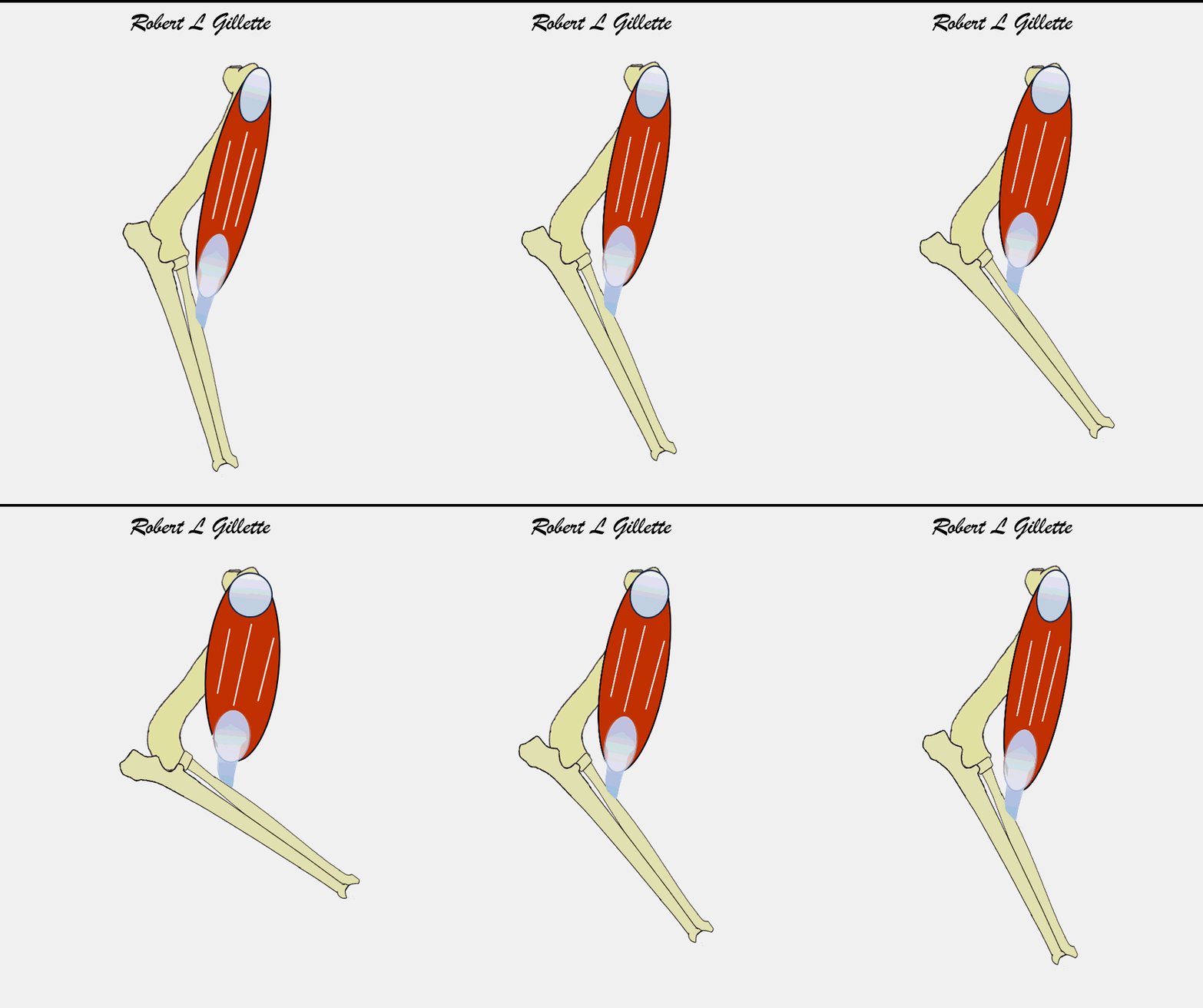
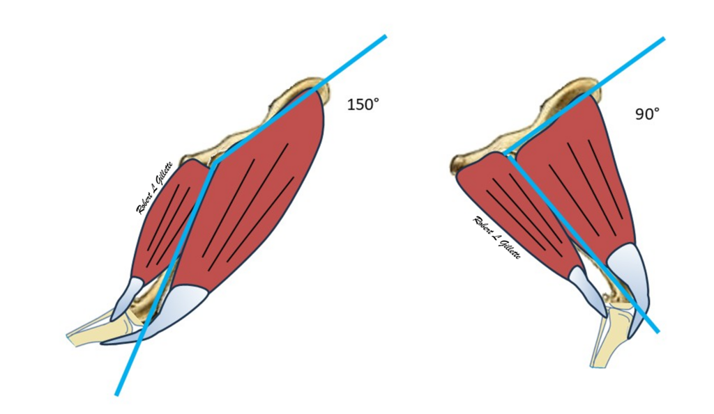
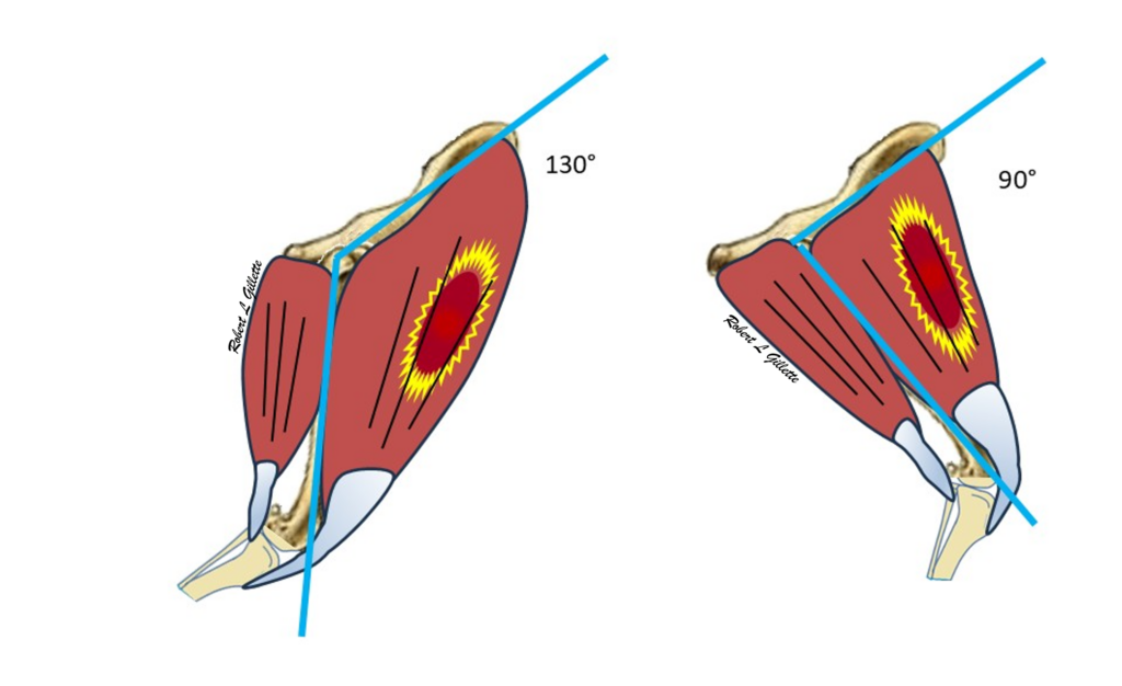
Muscles are the tissues that create the movements of the body. The nervous system works to coordinate these movements and bones are the structures that stabilize and support the body during these movements. Some movements are postural, in that they keep the body standing and stabilize how the body is postured. Other movements create the body’s actions. Locomotion, reaching and lifting are some action movements. The muscles create action around the articular joints of the bones. In the figure below, the quadriceps are located on the cranial (front side) of the femur or proximal rear limb. The hamstrings are located on the caudal (back side) of the femur proximal rear limb. In the example below, when the quadriceps muscle contracts, it flexes the hip joint or makes the joint angle smaller. When it does this, the hamstring muscles need to relax to allow the joint flexion. When the hamstrings contract the hip joint extends or the hip joint is greater. When this occurs, the quadriceps muscle needs to relax to allow hip extension. These are very general descriptions as the actions are much more complex than this. There is a balance of contraction/flexion of the opposing muscles which counterbalance each other for smooth movement.
The presence of a trigger point will change how these actions occurs. One common trigger point location is in the quadriceps. During hip flexion, the quadriceps may or may not be affected but typically the hip joint flexion will not appear to be affected. The trigger point will affect hip extension. The hamstrings will contract to extend the hip joint but the quadriceps will not fully relax. This impedes the ability of the hamstring muscles to extend the hip joint which results in a decrease in the angle of extension. These changes typically cannot be identified visually by the untrained eye but can be identified using video analysis and slow motion replay.
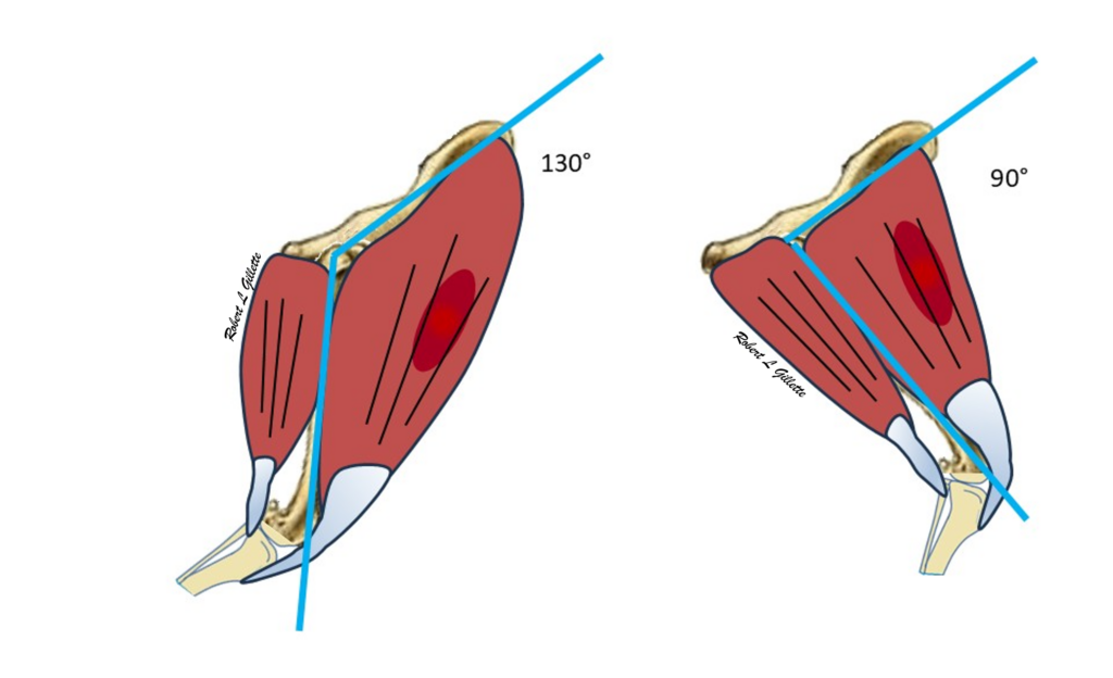
The other important note about trigger points, they are painful to the patient. The movements that occur when a trigger point is present can be painful but may not be described or demonstrated by the individual. This complicates any performance work or diagnostic process.
Movement Restrictors
- Some movements are postural, in that they keep the body standing and stabilize how the body is postured
- Other movements create the body’s actions
- Locomotion, reaching and lifting are some action movements
- The muscles create action around the articular joints of the bones
Movement Alterations and Mobility Analysis
- Identifying which areas are involved can be done using gait analysis and diagnostic palpation
- Video Analysis using slow motion playback can be very helpful to identify movement variants
Locomotion
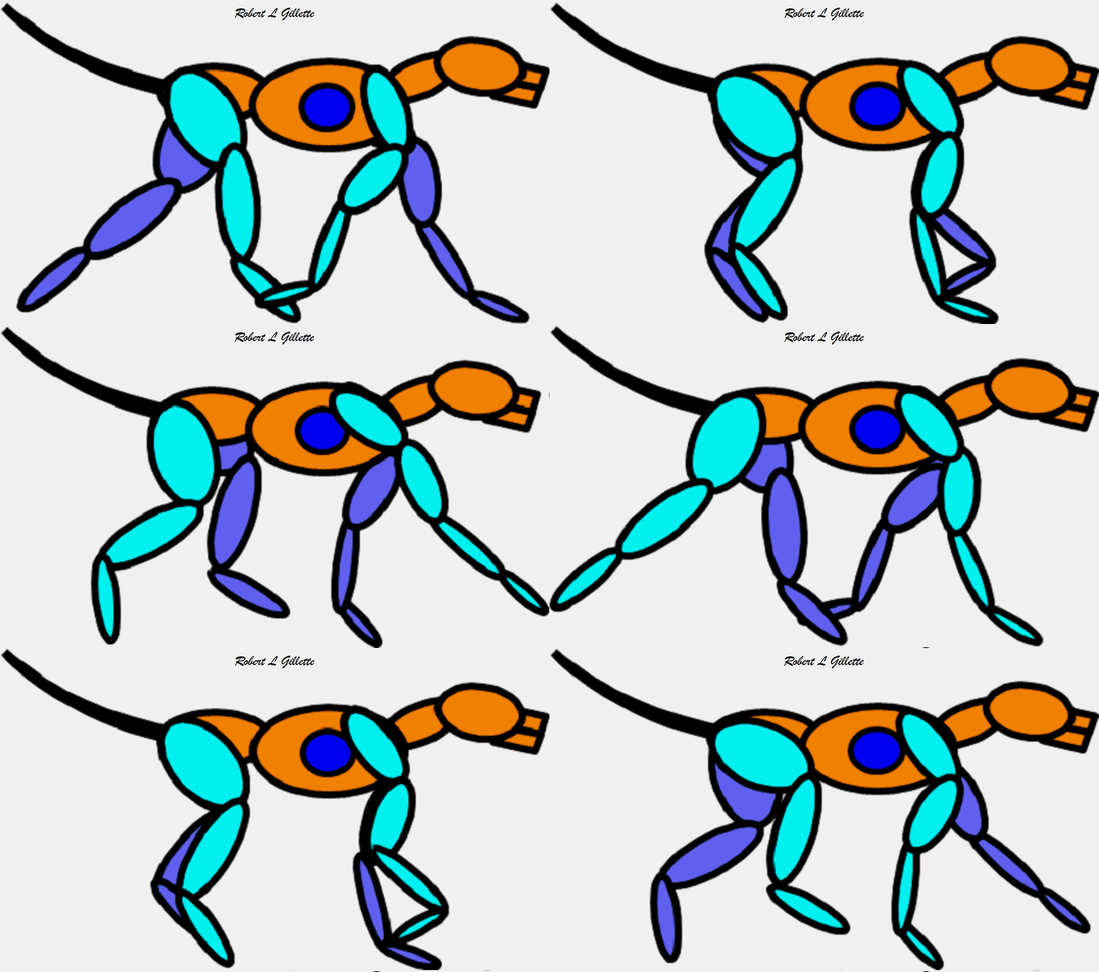
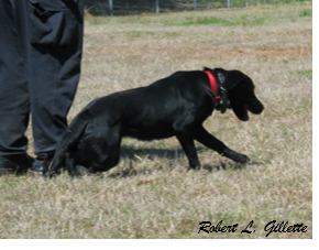
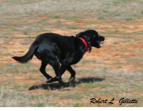
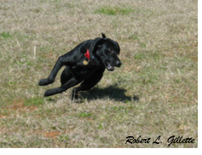
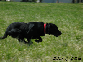
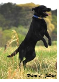
There are five variations of the normal locomotion. How these movements are affected can be an indicator of which groups are involved. Video analysis of the dog’s gait can be very helpful in diagnosing musculoskeletal issues.
|
 |
|
 |
|
 |
|
 |
|
 |
Functional Mobility
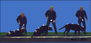
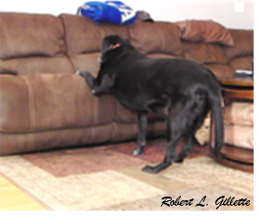
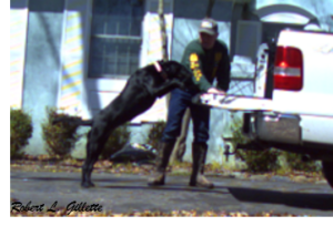
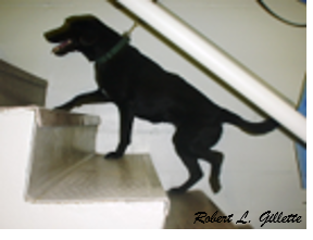
Functional Mobility is a term that can be used to describe actions involved in daily actions. Changes in functional mobility can be used to identify subtle musculoskeletal issues not diagnosed through gait analysis of the dog’s typical locomotory pattern. Video analysis of the dog’s functional movements can be very helpful in diagnosing these subtle musculoskeletal issues.
|
 |
|
 |
|
 |
|
 |
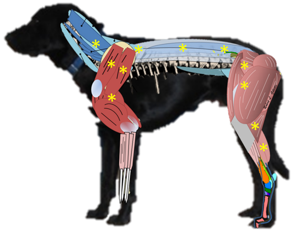
Common Locations that Trigger Points (TP) or Taut Muscle Bands (TMB) occur
Muscle trigger point diagnostics
The following information is offered only as an introduction to how muscle issues, and more specifically MTP’s, can affect movement in then dog. In any situation where an owner or handler perceives a problem in their dog, the first step should be to set up an appointment with their Veterinarian for discussion and a Veterinary workup.
- Neck mobility
- Reduced neck mobility
- Can be bilateral or unilateral
- Up & Down restrictions
- Left or right restrictions
- Reduce movement of shoulders
- Rule outs are vertebral, neural problems
- Radiographs
- Neural testing
- Once those are ruled out
- Consider muscle based issues
- Once other muscle issues are ruled out
- Trigger points, Taut Muscles Band
- Common TP locations
- Longissimus Cervicis, Transversospinalis
- Reduced neck mobility
- Front Limb mobility
- Typically unilateral but can be bilateral
- Reduce Shoulder Extension
- Pain on Flexion or Extension
- Rule outs are bone (Scapula, Humerus), joint complex (Shoulder), neural problems
- Radiographs
- Test for articular ligament damage
- Neural testing
- Once those are ruled out
- Consider muscle based issues
- Assess for muscle injury, tendon strain or muscle soreness
- Once other muscle issues are ruled out
- Trigger points, Taut Muscles Bands
- Common TP locations
- Triceps, Infraspinatus, Supraspinatus, Deltoids
- Typically unilateral but can be bilateral
- Thoracic Vertebral Segment
- Can be bilateral or unilateral
- Up & Down restrictions
- Left or right restrictions
- Restricted shoulder or hip movement
- Rule outs are vertebral, neural problems
- Radiographs
- Neural testing
- Once those are ruled out
- Consider muscle based issues
- Assess for muscle injury, tendon strain or muscle soreness (Check for weed awn migration)
- Once other muscle issues are ruled out
- Trigger points, Taut Muscles Bands
- Common TP locations
- Longissimus, Spinalis
- Can be bilateral or unilateral
- Lumbar Vertebral Segment
- Can be bilateral or unilateral
- Up & Down restrictions
- Left or right restrictions
- Restricted hip movement
- Rule outs are vertebral, neural problems
- Radiographs
- Neural testing
- Once those are ruled out
- Consider muscle based issues
- Assess for muscle injury, tendon strain or muscle soreness
- Once other muscle issues are ruled out
- Trigger points, Taut Muscles Bands
- Common TP locations
- Longissimus, Spinalis
- Can be bilateral or unilateral
- Rear Limb Mobility
- Typically unilateral but can be bilateral
- Reduce Hip Extension, Stifle Extension (common in CCL ruptures)
- Pain on Flexion or Extension
- Rule outs are bone (Pelvis, Femur, Tibia & Fibula), joint complex (Hip or Stifle), neural problems
- Radiographs
- Test for articular ligament damage
- Neural testing
- Once those are ruled out (although they are commonly associated with above R/O’s)
- Consider muscle based issues
- Assess for muscle injury, tendon strain or muscle soreness
- Once other muscle issues are ruled out
- Trigger points, Taut Muscles Bands
- Common TP locations
- Quadriceps, Tensor Fascia Lata, Sartorius, Gastrocnemius, Semimembranosus, Semitendinosus (the last three are typically associated with CCL ruptures)
- Typically unilateral but can be bilateral
Treatment & Management
- Treatment is based on the proper diagnosis and many times will involve concomitant etiologies or sources of issues
- This is especially true in the older patients
- Treatment for joint or bone pain will be dependent upon the source and may involve surgery or anti-inflammatory treatments
- Treatment for muscle sourced pain can also benefit from anti-inflammatory treatments
- Many times, releasing trigger points or taut muscle bands can be of great benefit for the patient or performance dog
The treatment and management regimen can be developed by the dog’s Veterinarian
- There are many therapeutic medical options & modalities that can help patients with these issues
- Please consult with a Veterinarian to determine the treatment protocols that are best suited for each dog’s case
- My historical experience, specific to trigger points and taut muscle bands, has changed over the past 30+ years of clinical practice as the technology has advanced
- Initially I used steroid injections in the TP site
- In the late 1990’s and 2000’s, I found I could use electrostimulation (E-stim or TENS)
- I chose this as it was noninvasive
- Treatment times were long
- Needed skin contact or a lot of gel for electrode connectivity
- In the 2010’s, I found I could use therapeutic laser to release the TP’s
- Treatment time was much shorter
- It didn’t need skin contact or gel
- Laser Class IV, III versus Class I
- Laser classes are based on power and potential for eye damage
- Class IV lasers have the potential for burning
- Class III are less likely for overheating
- Class I lasers have minimal chance of burning and are much safer for those individuals working in these areas of treatment
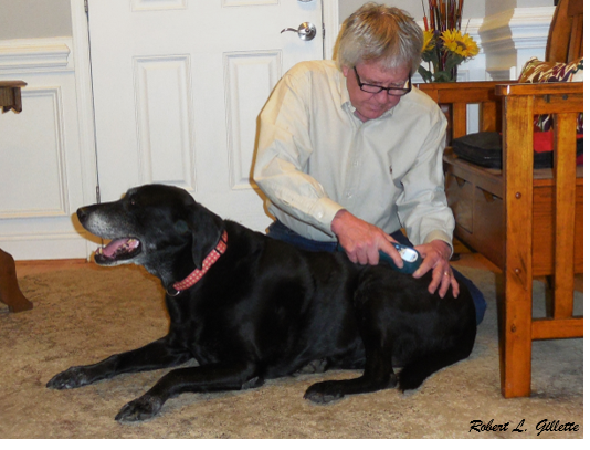
- I have been using a Class I laser since 2018 with a high level of success and minimal adverse effects
References
Gillette, R. L. (2020). Functional anatomy and biomechanics for the performance dog.
Guy, P. S., & Snow, D. H. (1981). Skeletal muscle fibre composition in the dog and its relationship to athletic ability. Research in Veterinary Science, 31(2), 244-248.
Osiak-Wicha, C., Kras, K., & Arciszewski, M. B. (2024). Comparative Analysis of Muscle Fibers in Selected Muscles of Working and Companion Dog Breeds. Animals, 14(24), 3576.
Kuzon Jr, W. M., Rosenblatt, J. D., Pynn, B. R., Marchetti, P. J., Plyley, M. J., & McKee, N. H. (1989). A comparative histochemical and morphometric study of canine skeletal muscle. Canadian Journal of Veterinary Research, 53(2), 125.
Brookhart, J. M., Parmeggiani, P. L., Petersen, W. A., & Stone, S. A. (1965). Postural stability in the dog. American Journal of Physiology-Legacy Content, 208(6), 1047-1057.
Ellis, R. G., Rankin, J. W., & Hutchinson, J. R. (2018). Limb kinematics, kinetics and muscle dynamics during the sit-to-stand transition in greyhounds. Frontiers in Bioengineering and Biotechnology, 6, 162.
Stark, H., Fischer, M. S., Hunt, A., Young, F., Quinn, R., & Andrada, E. (2021). A three-dimensional musculoskeletal model of the dog. Scientific reports, 11(1), 11335.
Brown, N. P., Bertocci, G. E., States, G. J., Levine, G. J., Levine, J. M., & Howland, D. R. (2020). Development of a canine rigid body musculoskeletal computer model to evaluate gait. Frontiers in Bioengineering and Biotechnology, 8, 150.
Gerwin, R. D. (2023). A new unified theory of trigger point formation: failure of pre-and post-synaptic feedback control mechanisms. International journal of molecular sciences, 24(9), 8142.
Gillette, Robert L. Athletic and Working Dog: Functional Anatomy and Biomechanics. RMG Enterprises, Inc., 2019.
Recent Comments